

Positron Emission Tomography (PET/CT)
PET/CT is a combination of two scan techniques – a PET Scan and a CT Scan. The combination of procedures can help to detect condition that may not show up on a standard X-ray, or a CT or PET scan alone. While a CT scan shows detailed anatomical structure, a PET scan provides information on organ function and metabolism. PET/CT is mainly used to detect various types of cancers and assessing cancer (staging, or restaging) that has spread (metastases), and for the evaluation of treatment response by looking at biochemical changes in the cancer, but there are growing clinical applications in cardiology and other fields.
PET imaging or a PET scan, is a diagnostic examination that involves taking physiologic images based on the detection of positrons, which are tiny particles emitted from a radioactive substance administered to the patient. The subsequent views of the human body developed by this technique are used to diagnose and/or evaluate a variety of diseases.
What are some common uses of the procedure?
- PET scans are used most often to detect cancer and to examine the effects of cancer therapy by characterizing biochemical changes in the cancer.
- These scans are performed on the whole body.
- PET scans of the heart can be used to determine blood flow to the heart muscle and help evaluate signs of coronary artery disease.
- PET scans of the heart can also be used to determine if areas of the heart that show decreased function are alive rather than scarred due to a prior heart attack, called a myocardial infarction.
- Combined with a myocardial perfusion study, PET scans differentiate non-functioning heart muscle from heart muscle that would benefit from a procedure, such as angioplasty or coronary artery bypass surgery, which would re-establish adequate blood flow and improve heart function.
- PET scans of the brain are used to evaluate patients who have memory disorders of an undetermined cause; who have suspected or proven brain tumors; or who have seizure disorders that are not responsive to medical therapy and, therefore, are candidates for surgery.
How should I prepare for the procedure?
- PET is usually done on an outpatient basis.
- Your doctor will give you detailed instructions on how to prepare for your examination.
- You should wear comfortable, loose-fitting clothes.
- You should not eat for four hours before the scan. You will be encouraged to drink water.
- Your doctor will instruct you regarding the use of medications before the test.
- Note: Diabetic patients should ask for any specific diet guidelines to control glucose levels during the day of the test.
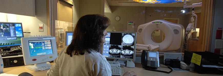
What does the equipment look like?
- You will be taken to an examination room that houses the PET scanner, which has a hole in the middle and looks like a large doughnut.
- Within this machine are multiple rings of detectors that record the emission of energy from the radioactive substance in your body and permit an image of your body to be obtained.
- While lying on a cushioned examination table, you will be moved into the hole of the machine.
- The images are displayed on the monitor of a nearby computer, which is similar in appearance to the personal computer you may have in your home.
How does the procedure work?
- Before the examination begins, a radioactive substance is produced in a machine called a cyclotron and attached, or tagged, to a natural body compound, most commonly glucose, but sometimes water or ammonia.
- Once this substance is administered to the patient, the radioactivity localizes in the appropriate areas of the body and is detected by the PET scanner.
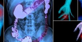
- Different colors or degrees of brightness on a PET image represent different levels of tissue or organ function. For example, because healthy tissue uses glucose for energy, it accumulates some of the tagged glucose, which will show up on the PET images.
- However, cancerous tissue, which uses more glucose than normal tissue, will absorb more of the substance and appear brighter than normal tissue on the PET images.
How is the procedure performed?
- A nurse or technologist will take you into a special PET examination room. You will lie down on an examination table and be given the radioactive substance as an intravenous injection (although, in some cases, it will be given through an existing intravenous line or inhaled as a gas).
- It will then take approximately 30 to 60 minutes for the substance to travel through your body and be absorbed by the tissue under study.
- During this time, you will be asked to rest quietly in a partially darkened room and to avoid significant movement or talking, which may alter the localization of the administered substance.
- After that time, scanning begins. This takes an additional 30 to 45 minutes.
- Some patients, specifically those with heart disease, may undergo a stress test in which PET scans are obtained while they are at rest, and again after undergoing the administration of a pharmaceutical to alter the blood flow to the heart. Usually, there are no restrictions on daily routine after the test, although you should drink plenty of fluids to flush the radioactive substance from your body.
What will I experience during the procedure?
- The administration of the radioactive substance will feel like a slight pinprick if given by intravenous injection.
- You will then be made as comfortable as possible on the examination table before you are positioned in the PET scanner for the test.
- You will be asked to remain still for the duration of the examination. Patients who are claustrophobic may feel some anxiety while positioned in the scanner. Also, some patients find it uncomfortable to hold one position for more than a few minutes.
- You will not feel anything related to the radioactivity of the substance in your body.
Who interprets the results and how do I get them?
- Patients undergo PET because their referring physician has recommended it.
- A radiologist who has specialized training in PET will interpret the images and forward a report to your referring physician.
- It usually takes one to three days to interpret, report, and deliver the results.
- You may be asked to bring any outside examinations with you, such as recent CT (CAT) scans or MRI scans.
What are the benefits vs. risks?
- Because PET allows study of body function, it can help physicians detect alterations in biochemical processes that suggest disease before changes in anatomy are apparent on other imaging tests such as CT or MRI scans.
- Because the radioactivity is very short-lived, your radiation exposure is extremely low. The substance amount is so small that it does not affect the normal processes of the body. The radioactive substance may expose radiation to the fetus of patients who are pregnant or the infants of women who are breast-feeding. The risk to the fetus or infant should be considered related to the potential information gain from the result of the PET examination. If you are pregnant you should inform the PET imaging staff before the examination is performed.
What are the limitations of Positron Emission Tomography?
- PET can give false results if a patient’s chemical balances are not normal.
- Specifically, test results of diabetic patients or patients who have eaten within several hours prior to the examination can be adversely affected because of blood sugar or blood insulin levels.
- Also, because the radioactive substance decays quickly and is effective for a short period of time, it must be produced in a laboratory near the PET scanner.
- It is important to be on time for the appointment and to receive the radioactive substance at the scheduled time.
- PET must be done by a radiologist who has specialized in nuclear medicine and has substantial experience with PET.
- The value of a PET scan is enhanced when it is part of a larger diagnostic work-up. This often entails comparison of the PET scan with other imaging studies such as CT or MRI.