

X-Ray
X-Ray
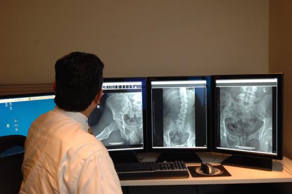
X-ray is a non-invasive exam that involves a low dose of radiation to produce pictures of the inside of the body, which make x-rays an extremely safe diagnostic test. X-rays are commonly used to determine broken bones (fractures) and damaged ligaments, and organ diseases such as pneumonia in the lung, or cancer.
Radiography, or an x-ray, as it is most commonly known, is the oldest and most frequently used form of medical imaging. Discovered more than a century ago, x-rays can produce diagnostic images of the human body on film or digitally on a computer screen. X-ray imaging is the fastest and easiest way for a physician to view and assess broken bones, such as skull fractures and spine injuries. At least two images (from different angles) are taken and often three images are needed if the problem is around a joint (knee, elbow or wrist). X-rays also play a key role in guiding orthopedic surgery and in the treatment of sports-related injuries.
X-ray may uncover more advanced forms of cancer in bones, although early screening for cancer findings requires other methods. To this end, radiologists have developed alternative imaging methods that do not rely on radiation, such as ultrasound and magnetic resonance imaging (MRI).
What Does the Equipment Look Like?
Radiography equipment consists of a large, flat table with a drawer that holds a tray into which an x-ray film cassette is placed. Suspended above the table is the apparatus that holds the x-ray tube that can be moved over the body to direct the x-ray.
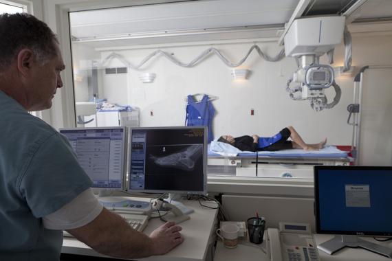
How does the procedure work?
- A technologist positions the patient on the examination table and places a film holder (cassette) under the table in the area of the body to be imaged.
- Sandbags or pillows may help the patient hold the proper position. Then the technologist steps behind a radiation barrier and asks the patient to hold very still without breathing for a few seconds.
- The radiographic equipment is activated, sending a beam of x-rays through the body to expose the film. The technologist then repositions the patient for another view, and the process is repeated.
- When x-rays are completed the patient will be asked to wait until the technologist checks the images for adequate exposure and motion.
- A radiologist who is a physician experienced in bone x-ray and all other types of radiology examinations will analyze the images, make a diagnosis and recommend treatment, such as casting in the case of simple fractures, or referral to other specialists, and send a signed report to the referring physician.
What are some common uses of the procedure?
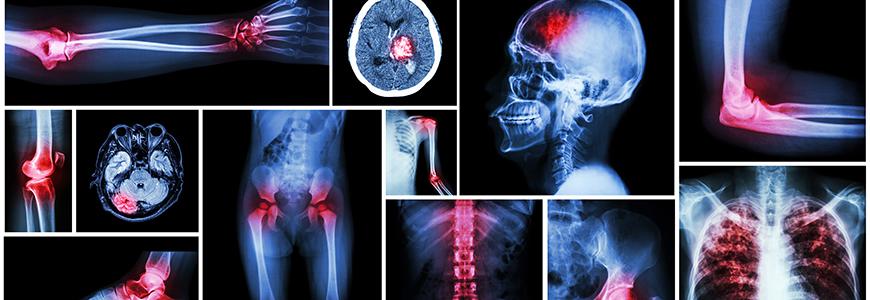
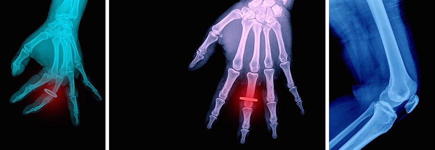
- Probably the most common use of bone radiographs is to assist the physician in identifying and treating fractures.
- X-ray images of the skull, spine, joints and extremities are performed every minute of every day in hospital emergency rooms, sports medicine centers, orthopedic clinics and physician offices.
- Images of the injury can show even very fine hairline fractures or bone chips, while images produced after treatment ensure that a fracture has been properly aligned and stabilized for healing.
- Bone x-rays are essential tools in orthopedic surgery, such as spinal repair, joint replacements or fracture reductions. X-ray images can be used to diagnose and monitor the progression of degenerative diseases such as arthritis.
- They also play an important role in the detection and diagnosis of cancer, although usually computed tomography (CT) or MRI is better at defining the extent and the nature of a suspected cancer.
- On regular x-rays severe osteoporosis can be visible, but bone density determination for early loss of bone mineral is usually done on specialized, more sensitive equipment.
How should I prepare for the procedure?
- There is no special preparation required for most bone radiographs.
- Once you arrive, you may be asked to change into a gown before your examination.
- You will also be asked to remove jewellery, eyeglasses and any metal objects that could show up on the images and overlap important findings.
- Women should always inform their doctor or x-ray technologist if there is any possibility that they are pregnant.
- You will be asked to remain still while the X-Ray is being taken
What will I experience during the x-ray procedure?
- X-ray imaging itself is painless. Some discomfort may result from lying on the table, a hard surface that may feel quite cold.
- Sometimes, to get a clear image of an injury such as a possible fracture, you may be asked to hold an uncomfortable position for a short time.
- Any movement could blur the image and make it necessary to repeat the procedure to get a useful, clear picture.
Who interprets the results and how do I get them?
- A radiologist is a physician experienced in bone x-ray and all other types of radiology examinations.
- The radiologist will analyze the images and send a signed report to your primary care or referring physician, who will inform you on your test results.
- New technology also allows for distribution of diagnostic reports and referral images over the Internet at many facilities.