

Magnetic Resonance Imaging (MRI)
Magnetic Resonance Imaging (MRI)
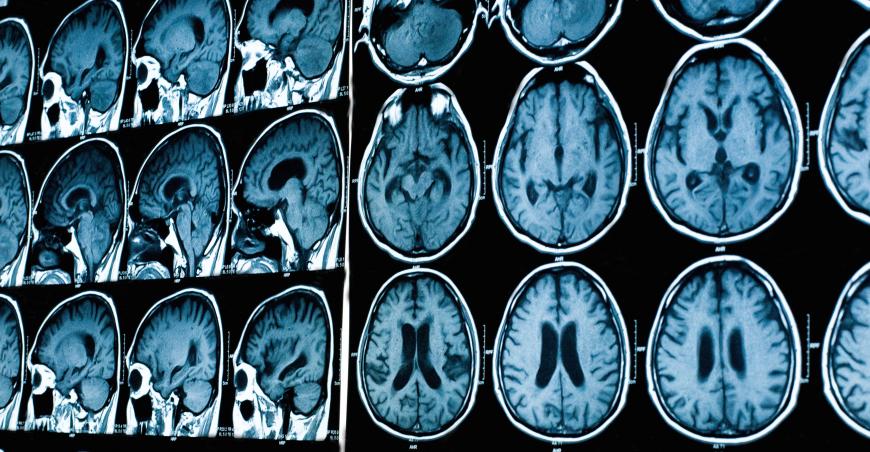
MRI is a non-invasive medical imaging exam that helps radiologists to diagnose a variety of diseases including cancer. MRI uses a powerful magnetic field, radio frequency pulses, and a computer to produce detailed pictures of organs, soft tissues, bone, and other structures inside the body. In some cases, contrast material may be used during the MRI scan to show certain structures more clearly. Unlike CT scanning or general x-ray studies, no ionizing radiation is involved with an MRI.
In many cases, MRI gives different information about structure in the body than can be seen with an X-ray, ultrasound, or computed tomography (CT) scan. MRI also may show problems that cannot be seen with other imaging methods. This information is then sent to a computer which processes all the signals and generates it into an image. The final product is a 3-D image representation of the area being examined. These images can then be displayed and examined on a computer monitor, transmitted electronically, printed, or copied to a CD.
In recent years patient-friendly units have been designed, and examination in such units is becoming increasingly available. These machines are both shorter and wider than a conventional MRI unit, and do not fully enclose the patient. Some of the newer C-shaped units are even open on all sides, and so are very attractive to patients who tend toward claustrophobia. A drawback is that image quality is not as consistently good.
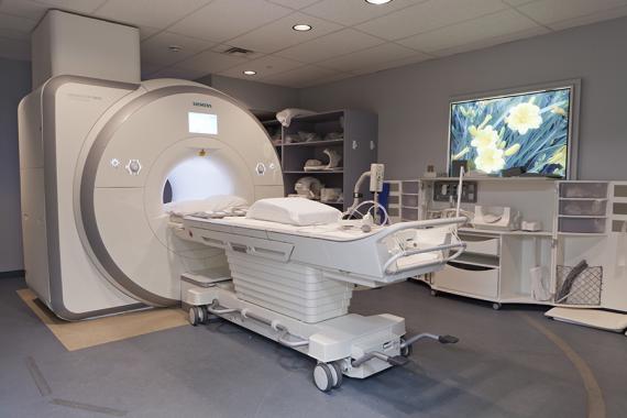
What Does the Equipment Look Like?
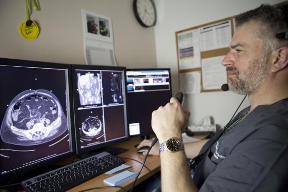
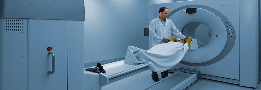
- An MRI machine looks like a long tube with a large circular section housing the magnet. Patients usually lie on their back on a table that is part of the MRI scanner.
- The head, chest and arms may be held in place with straps to help them stay still. It is very important to stay completely still while the scan is being done.
- The table is then slid into the round opening of the magnet. Then depending on where the MRI needs to be taken, the technician slides a coil to the specific area being imaged. The coil is the part of the machine that receives the MR signal.
- Inside the scanner patients will hear a fan and feel air moving. They will also hear loud tapping or snapping noises as the magnets are turned on and off throughout the exam as the MRI scans are done. People who get nervous in small-enclosed places may need medicine to help them relax while having an MRI scan.
Preparing for MRI
Preparation for your MRI will depend on the type of exam you are having.
Do I need to be fasting? Will I have to drink that awful stuff?
- If you are having a Cholangiogram (MRCP – Magnetic Resonance Cholangiopancreatography), you will have to fast for four hours prior to your exam.
- If you are having an MR-enterography exam, you will be required to drink oral contrast.
What should I wear?
- Gowns will be provided for your exam.
- You will be asked to remove ALL body piercings, jewelry, watches, eyeglasses, hairpins, wallets, and other metallic objects prior to your exam.
I am claustrophobic; will I be able to have a scan?
- Many scanners have short bore technology which makes for a more comfortable scanning experience.
- “Short bore” means that the length of the tube you’re in during your scan is shorter than usual. Unless you are having a scan of your head or neck, you may be able to see outside of the magnet.
- Discuss your claustrophobia with your physician. If sedation is prescribed by your doctor, bring it with you on the day of your exam. Inform the front desk staff upon arrival that you need to take prescribed medication for your exam.
Do I need to have my creatinine level performed before my exam?
- If your doctor has ordered an MRI with contrast (Gadolinium) and you have a history of diabetes, kidney (renal) issues, or hypertension, you’ll have to have your creatinine level obtained prior to your MRI.
- The blood test must be acquired within 6 weeks of your exam. The blood work must be performed within one week of the exam for all patients who have had or will be having a liver transplant.
What if I might be pregnant?
Please indicate any possibility of pregnancy to your physician and the scheduling office when you book the appointment. Inform the MRI technologist as well when you arrive at the department.
During my MRI
- After you have removed all metal objects, an MRI Technologist will position you on the table of the scanner.
- Your head will be placed in a padded plastic cradle or on a pillow, and the table will slide into the scanner.
- An intercom system will allow you and the technologist to be able to communicate with one another at all times.
- In order to obtain clear pictures, you will be asked to hold very still and relax. In some cases, you will be asked to hold your breath for up to 20 seconds.
- Any movement, especially of your head or back (even moving your jaw to talk) during the scan will blur and degrade the pictures. While the machine is taking your pictures, you will hear rapid, loud thumping noises coming from the scanner.
- During this time, you should breathe quietly and normally and refrain from any movement, coughing or wiggling. When the thumping noise stops, you must be still and maintain your position in the scanner. This noise comes from the gradient that allows the magnet to produce images.
- There will be multiple series of image acquisition, each with its own particular noises. The entire exam ordinarily takes between 30 and 60 minutes depending on exam type.
After your MRI
In most cases you will be able to leave at this point and resume regular diet and activities. There are occasional procedures, which will require that you stay and be monitored afterward. If you brought prior studies for comparison, these will be waiting for you at the front desk.
How will I know the results of my MRI?
After your MRI a subspecialized radiologist will read the images and dictate a report that will be sent immediately to your referring physician, who will contact you with the results.