
Nuclear Medicine
Nuclear medicine is a branch of medical imaging that uses small amounts of radioactive material to diagnose or treat a variety of diseases, including many types of cancers, heart disease, and certain other abnormalities within the body.
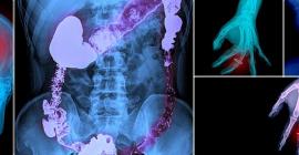
Nuclear medicine differs from other exams, such as x-ray, CT or MRI, because it images organ function, rather than just the anatomy. This means that it can show how an organ functions, not simply what it looks like. This allows us to not only monitor cancer, but also indicates the activity of many organs including the thyroid, heart, stomach and kidneys. Nuclear medicine is also well known for imaging the bones and joints to detect a number of abnormalities including trauma, fractures, arthritis or tumors.
What Does the Equipment Look Like?
- Nuclear medicine procedures are performed using either single photon emission computed tomography (SPECT) or positron emission tomography (PET).
- The gamma camera, which is encased in metal, is capable of detecting radiation and taking pictures from different angles.
- A gamma camera does not emit any radiation. It may be suspended over the examination table, or it may be beneath the table. Often, gamma cameras are dual-headed with one camera next to the other at a 90 degree angle.
- In some imaging centers, the gamma camera is located beneath the exam table and out of view. The camera may be located within a large, doughnut-shaped scanner similar in appearance to a computed tomography (CT) scanner. SPECT uses a gamma camera that rotates around the body to produce more detailed, three-dimensional images.
- A PET scanner is a large machine with a round, doughnut shaped hole in the middle, similar to a CT or MRI unit. Within this machine are multiple rings of detectors that record the emission of energy from the radiotracer in your body.
- A computer aids in creating the images from the data obtained by the camera or scanner.
How Does The Procedure Work?
- With ordinary x-ray examinations, an image is made by passing x-rays through the body from an outside source.
- In contrast, nuclear medicine procedures use a radioactive material called a radiopharmaceutical or radiotracer, which is injected into your bloodstream, swallowed or inhaled as a gas.
- This radioactive material accumulates in the organ or area of your body being examined, where it gives off a small amount of energy in the form of gamma rays.
- A gamma camera, PET scanner, or probe detects this energy and with the help of a computer creates pictures offering details on both the structure and function of organs and tissues in your body.
- Unlike other imaging techniques, nuclear medicine imaging exams focus on depicting physiologic processes within the body, such as rates of metabolism or levels of various other chemical activity, instead of showing anatomy and structure.
- Areas of greater intensity, called “hot spots,” indicate where large amounts of the radiotracer have accumulated and where there is a high level of chemical or metabolic activity. Less intense areas, or “cold spots,” indicate a smaller concentration of radiotracer and less chemical activity.
Exam Preparation
- Preparation for your Nuclear Medicine scan varies and instructions will be given to you when your exam is scheduled. Please bring previous imaging study results (x-ray, MRI, CT, etc.) such as reports, films or CD-ROMs if available.
- Notify the Nuclear Medicine staff if you are nursing or if there is a chance you could be pregnant.
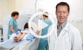
During the Exam
- A small and safe amount of radioactive “material/tracer” is used and goes to specific organs, bones, or tissues in the body.
- The tracer will be administered intravenously (IV injection, usually the arm) or orally; this is all contingent on the study obtained. It may take a few minutes, hours, or even a day or more for the tracer to reach the specific area to be studied. If there is a long wait period, you will be free to leave and return for your scan several hours later or the next day.
- Once you are positioned on a padded table, the tracer emits gamma rays that are detected by a gamma camera, which works with computers to form images that provide data about the body area in question.
- The amount of radiation needed for the exam is minimal and the body eliminates the material typically within 24 hours.
- You are encouraged to drink extra water to help remove the material more quickly.
After the Exam
- The images from your exam are diagnostically interpreted by a Nuclear Medicine Radiologist who prepares a diagnostic report, which is forwarded to hour physician.
- Your doctor will contact you with your results and discuss your results with you.