Ultrasound
Ultrasound is a simple, safe, painless diagnostic procedure that bounces high-frequency sound waves off parts of the body and captures the returning “echoes” as images. There is no injection or radiation exposure associated with ultrasound.There are many different types of ultrasound exams including abdomen, carotids, venous, pelvic, thyroid, renal, scrotum and obstetrical.
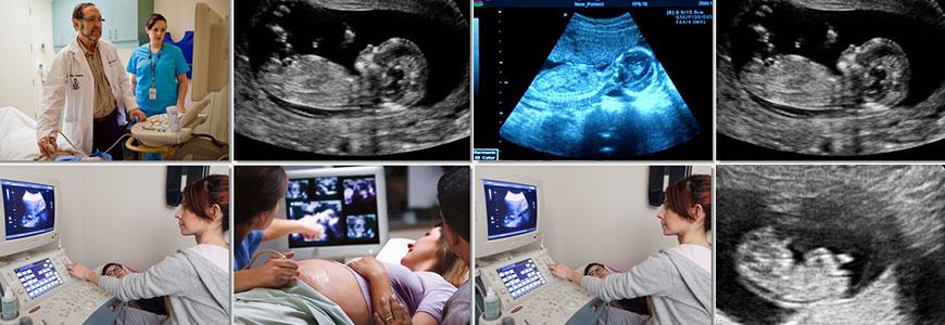
Ultrasound is able to capture moving images of pelvic and abdominal structures. When enhanced with a special technique called Doppler, ultrasound can also capture blood flow in the vessels and document moving images of the heart using echocardiography.
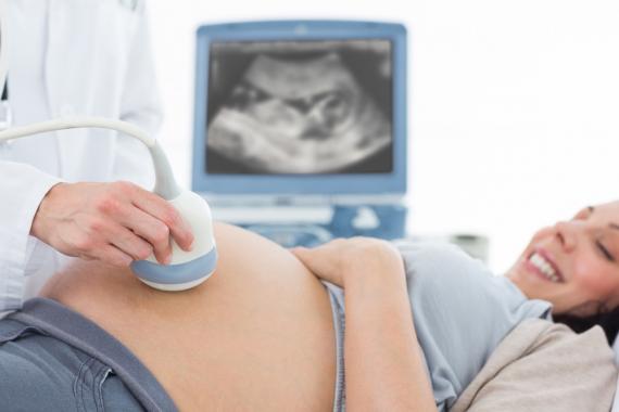
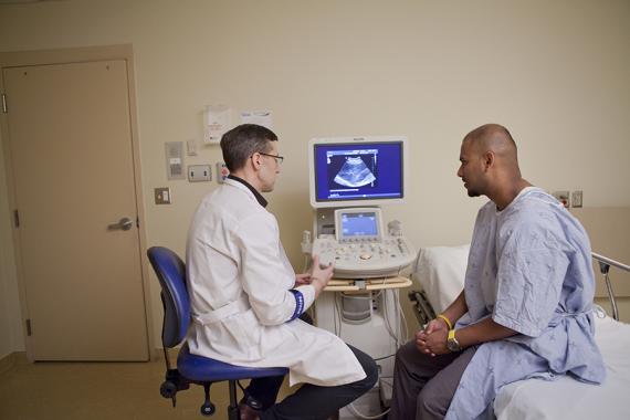
- The patient is positioned on a cushioned table and a clear water-based gel is applied to the skin in the area to be studied. The gel acts as a conductor.
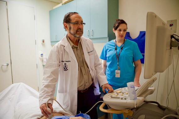
- A technologist, or radiologist (depending on the study) presses a transducer, a hand-held device that sends and receives ultrasound signals, firmly against the skin and sweeps it back and forth to image the area of interest.
- Images are instantly captured on a television-like monitor.
- The technologist transfers the images from your exam to the radiologist, a doctor who specializes in analyzing images of the specific area of the body you had examined. The radiologist will review the images, make a diagnosis and forward a report to the referring physician.
- Preparation for your ultrasound will depend on the type of exam; this will be reviewed with you when your appointment is scheduled. Wear comfortable clothing.
- Depending on your exam you may need to remove clothing and jewelry.
- Please bring previous imaging study results (X-ray, MRI, CT, etc.) such as reports, films or CD-ROMs if available.
- Most Ultrasounds are fast, easy, and painless.
- You will be positioned on a cushioned table and a clear water-based gel will be applied to your skin; the gel acts as a conductor.
- The technologist or radiologist presses a transducer, a hand-held device that sends and receives ultrasound signals, firmly against the skin and sweeps it back and forth to image the area of interest.
- Images instantly are captured on a television-like monitor and transferred to a radiologist to review and interpret. Depending on the type of exam, it can take anywhere from 20-60 minutes.
- The technologist transfers the images from your exam to the radiologist, who specializes in analyzing images of the specific area of the body you had examined.
- For instance if you had an ultrasound of your abdomen, an abdominal imaging radiologist will review your images.
- The radiologist will then dictate a diagnostic report, which upon completion is faxed, mailed or transmitted electronically to your physician.
- Your doctor will contact you with your results.