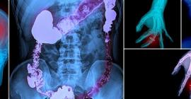


Virtual Colonoscopy
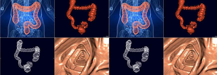
Virtual colonoscopy is a non-invasive procedure using sophisticated imaging techniques to generate a 3-D reconstruction of the inner surface of the colon, which can then be evaluated for abnormalities such as polyps by specially trained radiologists.
Virtual colonography is the latest development in colon cancer detection. Since it was first introduced in 1994, ongoing research has led to significant advances in the accuracy and usefulness of this procedure.
Unlike a traditional colonoscopy, virtual colonography is non-invasive and can be performed in a number of situations where traditional colonoscopy is contraindicated. It is also a good alternative for people who do not want to undergo traditional colonoscopy for a variety of reasons. The procedure makes use of sophisticated imaging techniques to generate a 3-D reconstruction of the inner surface of the colon, which can then be evaluated for abnormalities by specially trained radiologists.
CT colonography, also known as virtual colonoscopy, uses low dose radiation CT scanning to obtain an interior view of the colon (the large intestine) that is otherwise only seen with a more invasive procedure where an endoscope is inserted into the rectum and passed through the entire colon.
What Are Some Common Uses of the Procedure?
- The major reason for performing CT colonography is to screen for polyps, or cancers in the large intestine. Polyps are growths that arise from the inner lining of the intestine.
- A very small number of polyps may grow and turn into cancers.
- The goal of screening with CT colonography is to find these growths in their early stages, so that they can be removed before cancer has had a chance to develop.
What Does the Equipment Look Like?
- The CT scanner is a circular, ring-like machine with a large hole in the centre, like a doughnut.
- The patient lies still on a table that can move up or down, and slide into and out from the center of the ring. Within the machine, an x-ray tube on a rotating gantry moves around the patient’s body to produce the images, making clicking and whirring noises as the arm moves.
- The computer workstation that processes the imaging information is located in a separate control room, where the technologist operates the scanner and monitors your examination in direct visual contact and usually with the ability to hear and talk to you with the use of a speaker and microphone.
How should I prepare?
- You should wear comfortable, loose-fitting clothing to your exam. You will be given a gown to wear during the procedure. Women should always inform their physician and the CT technologist if there is any possibility that they may be pregnant.
- The bowel-cleansing regimen for CT colonography is similar to that for a colonoscopy. Your diet will be restricted to clear liquids the day before the examination. It is very important to clean out your colon the night before your CT colonography examination so that the radiologist can clearly see any polyps that might be present. You will be asked to take either a set of pills or a liquid laxative. Additional agents may also be taken the day before the exam. These may include small quantities of barium and iodinated liquids. These agents help the radiologist better distinguish stool from polyps by “tagging” the remaining stool and fluid.
- Be sure to inform your physician if you have heart, liver or kidney disease to be certain that the bowel prep will be safe. Your physician can advise you on dietary restrictions prior to the exam. You will be able to resume your usual diet immediately after the exam.
How does the procedure work?
- In many ways CT scanning works very much like other x-ray examinations. X-rays are a form of radiation—like light or radio waves—that can be directed at the body. Different body parts absorb the x-rays in varying degrees.
- In a conventional x-ray exam, a small burst of radiation is aimed at and passes through the body, recording an image on photographic film or a special image recording plate. Bones appear white on the x-ray; soft tissue shows up in shades of gray and air appears black.
- With CT scanning, numerous x-ray beams and a set of electronic x-ray detectors rotate around you, measuring the amount of radiation being absorbed throughout your body. At the same time, the examination table is moving through the scanner, so that the x-ray beam follows a spiral path. A special computer program processes this large volume of data to create two-dimensional cross-sectional images of your body, which are then displayed on a monitor. This technique is called helical or spiral CT.
- CT imaging is sometimes compared to looking into a loaf of bread by cutting the loaf into thin slices. When the image slices are reassembled by computer software, the result is a very detailed multidimensional view of the body’s interior.
- Refinements in detector technology allow new CT scanners to obtain multiple slices in a single rotation. These scanners, called multislice CT or multidetector CT, allow thinner slices to be obtained in a shorter period of time, resulting in more detail and additional view capabilities.
- Modern CT scanners are so fast that they can scan through large sections of the body in just a few seconds. Such speed is beneficial for all patients but especially children, the elderly and critically ill.
- For CT colonography, the computer generates a detailed 3-D model of the abdomen and pelvis, which the radiologist uses to view the bowel in a way that simulates traveling through the colon. This is why the procedure is often called a virtual colonoscopy. Two dimensional (2-D) images of the inside of the colon as well as the rest of the abdomen and pelvis are obtained and reviewed at the same time.
How is the procedure performed?
- The technologist begins by positioning you on the CT examination table, usually lying flat on your back or less commonly, on your side or on your stomach. Straps and pillows may be used to help you maintain the correct position and to help you remain still during the exam. Depending on the part of the body being scanned, you may be asked to raise your arms over your head.
- A very small, flexible tube will be passed two inches into your rectum to allow air to be gently pumped into the colon using a hand-held squeeze bulb. Sometimes an electronic pump is used to deliver carbon dioxide gas into the colon. Sometimes a small balloon is inflated on the rectal tube to help keep the tube positioned correctly. The purpose of the gas is to distend the colon as much as possible to eliminate any folds or wrinkles that might obscure polyps from the physician’s view.
- Next, the table will move through the scanner. Patients are asked to hold their breath for about 15 seconds or less before turning over and lying on their back or side for a second pass that is made through the scanner. In some centers the sequence of positions may be the opposite: facing upward first and then facing down. Once the scan is done, the tube is removed. The entire examination is usually completed within 15 minutes.
What will I experience during and after the procedure?
- The vast majority of patients who have CT colonography report a feeling of fullness when the colon is inflated during the exam, as if they need to pass gas. Significant pain is uncommon, occurring in fewer than 5 percent of patients. A muscle-relaxing drug may be injected intravenously or subcutaneously to lessen discomfort, but this is seldom necessary. The scanning procedure itself causes no pain or other symptoms.
- When you enter the CT scanner room, special light lines may be seen projected onto your body, and are used to ensure that you are properly positioned. With modern CT scanners, you will hear only slight buzzing, clicking and whirring sounds as the CT scanner revolves around you during the imaging process.
- You will be alone in the exam room during the CT scan, unless there are special circumstances. For example, sometimes a parent wearing a lead shield may stay in the room with their child. However, the technologist will always be able to see, hear and speak with you through a built-in intercom system. After a CT exam, you can return to your normal activities.
Who interprets the results and how do I get them?
- A radiologist with expertise in virtual colonography, will analyze the images and send a detailed report to the physician who referred you for the exam. The referring physician will discuss the results with you.
- In some cases, information about whether you have polyps is available immediately. Some imaging centers are equipped to perform colonoscopy and polyp removal the same day as the CT colonography.
- Follow-up examinations may be necessary, and your doctor will explain the reason why another exam is needed. Sometimes a follow-up exam is done because a suspicious or questionable finding needs clarification with additional views or a special imaging technique. A follow-up examination may be necessary so that any change in a known abnormality can be monitored over time. Follow-up examinations are sometimes the best way to see if treatment is working or if an abnormality is stable over time.
What are the benefits vs. risks?
Benefits
- This new minimally invasive test provides both 2-D and 3-D images that can depict many polyps and other lesions as clearly as when they are directly seen by conventional colonoscopy.
- CT colonography has a markedly lower risk of perforating the colon than conventional colonoscopy. Most people who undergo CT colonography do not have polyps, and can be spared having to undergo a full colonoscopy.
- CT colonography is an excellent alternative for patients who have clinical factors that increase the risk of complications from colonoscopy, such as treatment with a blood thinner or a severe breathing problem.
- Elderly patients, especially those who are frail or ill, will tolerate CT colonography better than conventional colonoscopy.
- CT colonography can be helpful when colonoscopy cannot be completed because the bowel is narrowed or obstructed for any reason, such as by a large tumor.
- If conventional colonoscopy cannot reach the full length of the colon—which occurs up to 10 percent of the time—CT colonography can be performed on the same day because the colon has already been cleansed.
- CT colonography provides clearer and more detailed images than a conventional barium enema x-ray examination.
- CT colonography can detect abnormalities outside of the colon, including early-stage malignancies and potentially dangerous conditions, such as abdominal aortic aneurysms.
- CT colonography is tolerated well. Sedation and pain relievers are not needed, so there is no recovery period.
- CT colonography is less costly than colonoscopy.
- No radiation remains in a patient’s body after a CT examination.
- X-rays used in standard CT scans have no immediate side effects.
Risks
- There is a very small risk that inflating the colon with air could injure or perforate the bowel.
- This has been estimated to happen in fewer than one in 10,000 patients.
- There is always a slight chance of cancer from excessive exposure to radiation. However, the benefit of an accurate diagnosis will generally outweigh the risk.
- The effective radiation dose for this procedure varies.
- Women should always inform their physician and x-ray or CT technologist if there is any possibility that they are pregnant. CT scanning is, in general, not recommended for pregnant women unless medically necessary because of potential risk to the baby.
What are the limitations of CT Colonography?
- A person who is very large may not fit into the opening of a conventional CT scanner or may be over the weight limit—usually 450 pounds—for the moving table. For very large patients, some facilities have extra-large bariatric-capable CT scanners. If this is necessary, contact your doctor for more information.
- CT colonography is strictly a diagnostic procedure. If any clinically significant polyps are found, they will have to be removed by conventional colonoscopy.
- The ability of CT colonography to differentiate stool from artifacts and smaller polyps may not be as good as that of conventional colonoscopy.
- CT colonography is not recommended for patients who have active Crohn’s disease, ulcerative colitis, inflammatory bowel disease or diverticulitis, because of increased risk of perforating the colon. Patients with a history of bowel perforation and those experiencing severe pain or cramps on the day of the examination should not undergo CT colonography.